Description
Props and Materials
Concepts
Learning Objectives
Set-Up
How To Demonstrate
Questions To Ask
Sample Dialogue
Background Information
Credits
For a paper copy of this guide, go here.
Description
This activity will explore a drug currently being tested by local scientists that causes brain tumors resulting from a particular type of cancer to glow, allowing surgeons to easily see the tumor and remove it without damaging healthy brain cells. In the activity, visitors will attempt to remove a tumor from a model brain, realizing the difficulty of identifying tumor cells. As they brainstorm a solution to this problem, they will learn via a magnet activity that a molecule in scorpion venom can attach to tumor cells. They will mark the venom with invisible ink markers, then return to a model brain with a blacklight to reveal that certain parts glow, allowing for easy removal of the tumor.
(back to top)
Props and Materials
Permanent______________________________________
|
 |
|
 |
|
 |
- Treat all props with respect.
- The black and white brain blocks could easily become lost. Please maintain control of them.
- While not sharp, the giant tweezers could easily poke and hurt someone. Please maintain control of them.
- Invisible ink markers are fun. Too much fun. While visitors can draw anything they want on their sticky notes, they should not draw on other things or each other. When visitors are done using the markers, put them away as soon as possible.
- Black lights are fun. Too much fun. While visitors can shine the light on themselves and each other to see how it works, they should not shine it in each others' eyes or run away with it to start a rave. Please maintain control of it. And them.
Concepts
- While surgery is currently one of the most effective ways to eliminate cancer, it remains dangerous due to the fact that tumor cells are often indistinguishable from healthy cells.
- Local scientists are working on a drug known as "tumor paint" that causes tumor cells resulting from a particular kind of cancer to glow, allowing surgeons to more easily identify tumor cells and remove them without harming healthy tissue.
Learning Objectives
- Looking a model of a brain, visitors will attempt to identify a tumor. Looking at a two-dimensional puzzle of a brain, visitors will attempt to remove a tumor, leading them to realize tumor cells may look exactly like healthy cells.
- Visitors will remove tiles from a box with a scorpion on it, representing removing venom from a scorpion. They will discover that some of these tiles stick to other tiles representing brain cells, helping them to visualize that venom can attach to tumor cells. They will then use invisible ink markers to draw on the tiles, representing the way scientists attach fluorescence to the venom to make the tumor glow. Returning to the original brain models, visitors will discover with a black light that certain portions glow, allowing them to easily identify tumor cells.
Set-Up
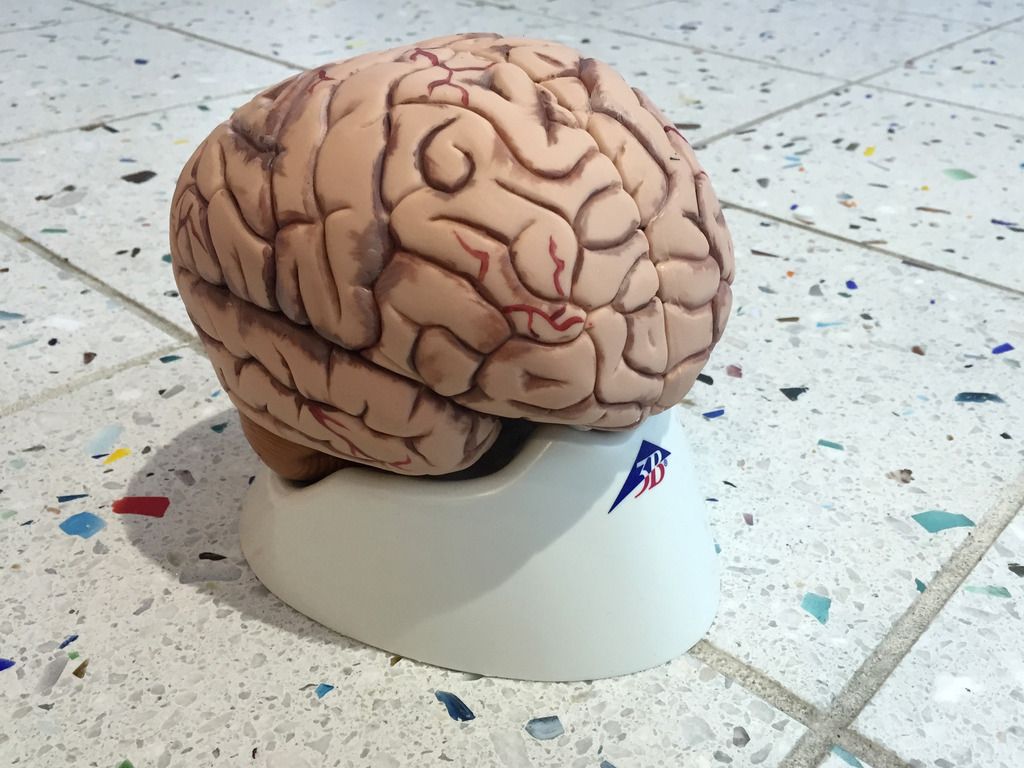
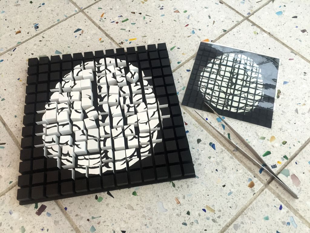
- Take out the the black brain board with the black-and-white blocks in the middle, the giant tweezers and the model of the brain. The black-and-white blocks don't need to be in any particular arrangement, but if you have time or want it looking nice, you can use the laminated brain picture to arrange the blocks.
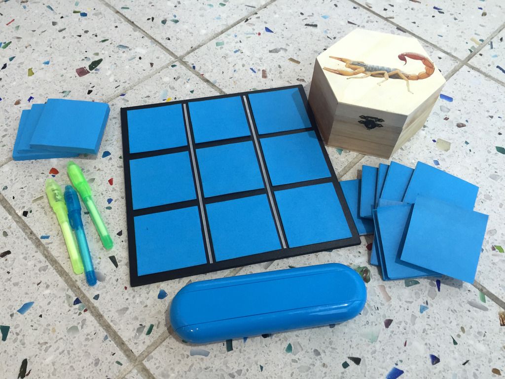
- Put blank sticky notes on the magnetic tiles, and put the tiles in the scorpion box.
- Have the scorpion box, blue brain cell board, invisible ink markers and black light ready.
How To Demonstrate
- Attract visitors to your activity by asking them questions like, "Do you want to be a brain surgeon?" or, "Do you want to help current researchers cure cancer?"
- Show them the brain model, letting them know that the brain has cancer. Ask them if they know what cancer is, letting them know that it has to do with cells. Cells are the building blocks of the body, and cancer causes cells to grow out of control, resulting in tumors. Ask them if they can find the tumor on the brain.
- Show them the black brain board with the black-and-white brain blocks. Let them know that a current way to treat cancer is surgery, which involves cutting out the tumor. Giving them the giant tweezers, challenge them to become a brain surgeon by removing the blocks they think might be a tumor. Once they remove some blocks, ask them whether they are sure they got all the cancer out. Discuss the function of the brain and ask them if they think they removed any healthy brain pieces, getting them to think about what can happen if healthy pieces get removed. Let them know this is something brain surgeons trying to remove cancer face every day.
- Take out the blue brain cell board, letting the visitor know that each tile is a brain cell and some of these cells are cancer, but some aren't. Ask again if they can tell which one belongs to a tumor and which is healthy. Take out the scorpion box, letting them know that venom from the deathstalker scorpion reacts with tumor cells of certain cancer. Have them remove the tiles from the scorpion box and find the off-white edges on the tiles. Have them line up the off-white edges with the off-white lines next to the blue squares on the board. They will notice that some of the venom tiles stick to some of the brain cells. Let them know that this venom attaches to tumor cells but not healthy cells.
- Ask the visitor if they can now identify which cells on the blue brain cell board are cancerous, pointing out that even though the venom is attached, it is difficult to tell which cells have venom attached by just looking at it. Let the visitor know that if we marked the venom in some way--for instance, by making the venom light up--we could more easily see the cancer cells. Give the visitors the invisible ink markers, telling them to mark the venom. Put the invisible ink markers away and give them the black light, showing them that now the venom--and thus the cancer cells--are easily visible.
- Have the visitor put the black-and-white brain blocks back on the black brain board. Use the black light to illuminate the board, and have them try once more to remove the tumor. Ask them the same questions as before--are they sure they got it all? Did they remove any healthy brain pieces? Have them shine the light on the brain model as well, letting them know that this procedure is something scientists are currently testing. They are able to synthesize one of the chemicals from the venom of the deathstalker scorpion, then add a tiny bit of fluorescence so that surgeons can see the venom chemical, which attaches to certain kinds of tumor cells. This drug, known as "tumor paint", has been tested on mice, and is currently being tried with human patients.
Questions To Ask
- What does your brain do?
- Do you know what cancer is?
- What are some ways we currently treat cancer?
- What might make surgery on a tumor difficult?
- What could we do in order to more easily identify tumors?
- What would happen if we removed a healthy piece of brain?
- Why would scientists want to make tumors glow?
- Do you think tumor paint will be used more in the future? Why or why not?
Sample Dialogue
Key:
- P Presenter
- G Guest
- Bold italics indicate action.
- Italics indicate a note to the presenter.
- □ indicates a cue
| P | Hi there! Want to be a brain surgeon with me? | |
| G | Okay! | |
| P | Do you know what this is? | |
| G | A brain. | |
| P | You're right. This particular brain has cancer. Do you know what cancer is? | |
| G | It's bad and it killed my great aunt. | |
| P | I'm really sorry about that. Cancer affects a lot of people. Do you want to talk about this? | |
| G | Sure. | |
| P | Okay. Cancer is about cells. Do you know what cells are? | |
| G | Like . . . the things that make you who you are. | |
| P | Right. Cells are little building blocks that make up different tissues, and different tissues make up our organs, like our heart and muscles and skin and eyes--as well as our brain. Cancer makes the cells in those organs grow out of control, which creates tumors. Have you heard of tumors? | |
| G | Yeah. They are also bad. | |
| P | Right. Some tumors will not hurt you, but other tumors are a result of cancer, which can definitely hurt you. This brain has a tumor--can you see it? | |
| G | Um, maybe. | |
| P | Cool. Do you know how we treat cancer? | |
| G | You take a lot of drugs and your hair falls out. | |
| P | Chemotherapy is one way of treating cancer. You take some chemicals and they work like a poison on the cancer cells. Another way is radiation, which is like burning the cancer out. But we also use surgery, which we can use to cut the cancer out. Do you want to do surgery on a brain? Don't worry, it's not real. | |
| G | Okay. | |
| P | Great! This black-and-white brain has pieces you can remove. Your job is to remove the tumor using these tweezers. | |
| G | It all looks the same. | |
| P | You're right! This is really hard. You might have to guess, which is something surgeons try very hard not to do. They want to make sure they get the tumor and not healthy brain. Go ahead and try it. | |
| G | Okay. | |
| P | Do you think you got it all? | |
| G | I don't know. | |
| P | What do you think would happen if a surgeon left part of the cancer in the brain? | |
| G | They'd still have cancer? | |
| P | Right. Okay, what do you think would happen if a surgeon accidentally removed a healthy part of the brain? | |
| G | I don't know. | |
| P | What does your brain do? Do you know why you have one? | |
| G | It's to think and stuff. | |
| P | Yeah. So what if part of a healthy brain got taken out? | |
| G | You wouldn't be able to think? | |
| P | Maybe. Maybe they just wouldn't remember everything, or they wouldn't be able to talk like they used to. Does that sound like a good thing? | |
| G | No. | |
| P | Okay, so we want to avoid removing healthy brain. Scientists are looking for a way to mark the tumor cells so they can identify the pieces we want to take out and the pieces we want to keep. Can you think of a way to do that? | |
| G | Like maybe the tumor could be a different color. | |
| P | Great idea. But how do we know which part is a tumor so we can color it? | |
| G | I don't know. | |
| P | That's okay. In order to find the tumor cells, scientists have started using this scorpion. Do you know what a scorpion is? | |
| G | It's like a thing that lives in the desert. And it can sting you. With its tail! | |
| P | Right! Scorpions can definitely sting you with venom. But this scorpion--the deathstalker scorpion--has special venom. It reacts with cancer cells. These blue tiles here are brain cells. Some of them have cancer; some don't. Want to take some venom out of the scorpion and see what it does to the brain cells? | |
| G | Sure. | |
| P | Great. Open the scorpion box and take out some venom. | |
| G | Um . . . this is not venom. These are just more tiles. | |
| P | You're right. This is just a representation of what the venom does with cancer cells. Can you find the white lines on the sides of these venom tiles? | |
| G | Sure. | |
| P | Great! Line up those white lines with the lines on this board, putting the venom tiles on top of the brain cell tiles. What is happening with some of the tiles? | |
| G | They stick. | |
| P | Right. Does the venom stick to all the brain cells? | |
| G | No. | |
| P | It only sticks to certain brain cells. Why might some of these cells be different than others? | |
| G | They have cancer? | |
| P | Right. Now we have something that points out which cells have cancer . . . but is it easy to see? | |
| G | Yes? | |
| P | But it's all the same color, right? We were going to try making the tumor a different color than the rest of the brain. What should we do so we know where the cancer cells are? | |
| G | Maybe color them? | |
| P | Right. If we color the venom, then when the venom sticks to the cancer cells, the cancer cells will be colored too. For this, scientists use something that glows, so that surgeons can easily see the cancer. I'm going to take all the venom off again, and then we can color them with something that glows as well. Here, use this marker to color the venom. | |
| G | Yay! Invisible ink markers! | |
| P | You can use it to color all of the venom if you want! | |
| G | I'm going to draw a flower and a pony and a dragon and Alexander Hamilton! | |
| P | Good job. Now try putting the venom back on the brain cells and take off the ones that don't stick. | |
| G | Okay. | |
| P | We still can't see the cancer cells very well, but since we used those markers, we can see it under a black light. Here, try it. | |
| G | Oh my God a black light! The panda on my shirt is glowing! My shoelaces are glowing! Alexander Hamilton is glowing! I'm going to go color all the sea stars in the tide pool and make them all glow! | |
| P | Oops, I forgot to put the markers away. Okay, now we can see the cancer cells, right? | |
| G | And Alexander Hamilton. | |
| P | Great. Do you think this would make brain surgery on a cancerous tumor easier? | |
| G | Yes? | |
| P | Let's try it! Let's put the blocks back that you took out, then shine the black light on the brain while you perform surgery. | |
| G | Wow, I was way off on this tumor thing. | |
| P | That's okay. Is it easier now you can see where the tumor is? | |
| G | Yeah. | |
| P | Do you think this would work on a real brain? | |
| G | Maybe. | |
| P | How about you shine the black light on the model of the real brain? | |
| G | I can see the tumors. | |
| P | Great. Scientists are trying this right now. When they found that something in the scorpion's venom attached to certain cancer tumors, they starting making that chemical from the venom. Then they attach just a little bit of fluorescence to the venom, so that when the venom attaches to the tumor, the tumor glows. Surgeons can more easily see where the tumor is when they go to remove it. Do you think this is a good idea? | |
| G | You mean I could literally put Alexander Hamilton on my brain. | |
| P | Not quite. But you could make a little bit of someone's brain glow if they had cancer. | |
| G | Seems legit. | |
| P | The chemical from the venom with the bit of fluorescence attached is called tumor paint, and local scientists are testing it right now. Do you think this would be helpful to deal with cancer in the future? | |
| G | Yes. But it would probably be better if they just got rid of it so that you didn't have to do surgery. | |
| P | That's a good point. There are a lot of different things scientists are doing to deal with cancer, and this is just one of them. You can find out more about it in the exhibit behind me. | |
| G | I want to draw George Washington with the glow-in-the-dark markers. | |
| P | Thank you for looking at these brains with me! |
Background Information
Click to jump to any of these topics:
Cell Proliferation
Cancer
Cancer Treatment
Tumor Paint
Glioma
Optimized Peptides
Cell Proliferation__________________________
In order to understand cancer, it is necessary to first understand cell proliferation. Many cell signals are involved in determining which cells should divide and when they should divide. Cells receive “DIVIDE!” signals only when the organism needs to undergo growth, development, reproduction, or if there is a need to repair damaged tissues. Cells receive “DON’T DIVIDE!” signals if there is DNA damage, chromosomal abnormalities, no need for new cells, or if the cells are mature and specialized.
There are both internal and external “DIVIDE!” and “DON’T DIVIDE!” signals. Internal signals consist of proteins that monitor conditions in the cell and prevent it from dividing if no “DIVIDE!” signal has been received or if something is wrong. If something IS wrong, proteins trigger either DNA repair machinery or apoptosis (programmed cell death). External signals can also tell cells whether the environment is right for division, which tells them whether to “DIVIDE!” or “DON’T DIVIDE!” Normal cells have to receive external “DIVIDE!” signals before they divide, and they also heed “DON’T DIVIDE!” signals.
(back to topic list)
Cancer__________________________
According to the National Cancer Institute, about 40% of people will be diagnosed with some type of cancer at some point during their lifetime. In 2012, there were an estimated 13.8 million people living with cancer just in the United States. The prevalence of cancer makes the study of the disease and the search for better treatments important public health issues.
Cancer is, simply put, out-of-control cell growth. Cancer cells are cells with mutated genes that no longer respond normally to cell signals and cell cycle control systems. They may divide in the absence of “DIVIDE!” signals, and/or they may ignore “DON’T DIVIDE!” signals.
Many cancers are caused by mutations in cell division regulation genes; these genes make the internal “DIVIDE!” and “DON’T DIVIDE!” signals. Other types of mutations that may lead to cancer include mutations in genes that regulate the expression of the cell division regulation genes, genes that are involved in DNA damage repair, genes that control apoptosis, and chromosomal abnormalities that may affect multiple genes.
So what causes the mutations in these genes? Mutations may be inherited, mutations may occur during DNA replication if errors are not corrected, or mutations may be caused by environmental mutagens. A strong causal link has been established between smoking and lung cancer. Ultraviolet, X-ray, and gamma-ray radiation have also been shown to cause mutations that can lead to cancer. Other environmental factors have been implicated, but the data mainly show correlations rather than strong causal links: pollutants, pesticides, alcohol, and char-grilled or fried meats. Certain viruses (hepatitis B and C, HIV, HPV, HTLV) may increase the risk of cancer.
Mutations occur randomly in cells due to environmental factors or mistakes made during DNA replication. Most mutations are fixed by repair machinery. Occasionally, the mutations persist, leading to cell death. If the mutated cell somehow escapes apoptosis, then it may continue to live and accumulate further mutations.
Cancer is a multi-step process. Many mutations in multiple genes have to accumulate before a cell can bypass all of the regulations and signals needed in order to divide in an out-of-control manner. Precancerous cells slowly become cancerous as they accumulate mutations; this accumulation of mutations may take decades depending on how many mutations were initially inherited and the quantity of environmental mutagen exposure. Mutations that are inherited would be present in all of the cells of a person’s body and may increase that individual’s risk of cancer due to the fact that the individual has fewer mutations to accumulate in his/her cells before cancer develops.
(back to topic list)
Cancer Treatment__________________________
Currently, the three most common methods of treating cancer are surgery, radiation, and chemotherapy.
The aim of surgery is to cut out any and all cancer cells, so that they may not continue to divide and affect the patient. The problems with any type of surgery include the risk of the surgeon damaging surrounding tissues, the patient suffering an adverse reaction to the anesthesia, and the risk of surgical wound infection. Surgery to remove cancerous growths has additional risks. The surgeon may not be able to identify, observe, or remove all of the cancer cells present, so the patient may experience a recurrence of the cancer. The cancer may have spread with other undetected tumors away from the surgical site or cancer cells circulating in the bloodstream or lymphatic system of the patient.
Radiation therapy is used as a cancer treatment to severely damage DNA so that the cancer cells are no longer able to divide. Although the radiation is localized as much as possible to the site of the tumor, the radiation may damage the DNA of the surrounding non-cancerous cells; this may cause mutations that lead to other cancers in the future.
Cancer cells divide more rapidly and more often than normal cells, so chemotherapy exploits this fact by utilizing drugs that prevent cell division. Unfortunately, chemotherapy drugs also affect the rapidly dividing normal cells of the body, including skin cells, blood cells, and the cells lining the gastrointestinal tract. This accounts for the unpleasant side effects of chemotherapy. Drugs that block cell cycle progression or angiogenesis (the formation of blood vessels going to and from a tumor) may be used in chemotherapy. Taxol, a compound isolated from yew trees in the 1950s, was found to freeze the cytoskeleton so that cell division cannot occur.
Current cancer research is exploring other treatment methods. DNA microarray analysis reveals which genes are turned on or off in a particular tumor. This can determine the unique genetic “fingerprint” of the tumor, enabling individualized treatment tailored to each patient’s unique needs and exploits each tumor’s unique weaknesses. Pedigree analysis of families with a high incidence of cancer at young age is being used to determine specific genes involved in cancer. Studying the functions of these genes may lead to other avenues of treatment.
(back to topic list)
Tumor Paint __________________________
Surgery to remove brain cancer is especially difficult. The cancer cells often look very similar to normal cells, and only the surgeon’s experience and expertise can be used to identify the cancer cells. If the surgeon doesn’t remove all of the cancer cells, the cancer may return. If the surgeon removes too much tissue, irreversible brain damage may occur. Brain tumor surgery would be easier if there was a way to visualize the cancer cells.
Dr. Jim Olson’s laboratory at the Fred Hutchinson Cancer Research Center specializes in finding and developing therapeutics for brain cancer in children. Many years ago, Olson became obsessed with the question: “Would it be possible to light up a cancer cell?” A few years ago, Olson decided to revisit this question and asked one of his researchers, Dr. Patrick Gabikian, to search published literature for something that binds to brain cancer cells. Gabikian found one promising lead: chlorotoxin from death stalker scorpion venom binds to certain cancer cells in brain tumors known as gliomas.
Olson’s lab took chlorotoxin and tagged it with Cyanine5.5 NHS ester, a commercially available fluorescent dye used to label parts of proteins known as peptides. This created a molecule that specifically binds to glioma cells and labels those cells with fluorescence.
This tumor paint (official name: BLZ-100) can be used in real time during surgery. It crosses the blood-brain barrier, meaning that it can be injected into the blood and the fluids around the brain will not filter it out, which allows it to reach the brain and attach specifically to cancer cells. From 2005 to 2011, preclinical trials with mice and dogs found that tumor paint was 5,000 times more sensitive than an MRI in detecting cancer. It can make a group of as few as 200-cancer cells glow.
The first clinical trial of tumor paint began on May 25, 2015 at Seattle Children’s Hospital. The trial is run by Blaze Bioscience Inc. and is in phase 1 (testing for safety and most effective schedule and dose). During this trial, up to 27 people (from infants to young adults under the age of 30) diagnosed with a brain tumor will have their operations performed using tumor paint.
(back to topic list)
Glioma __________________________
Gliomas are tumors that form in the glial cells of the brain. Glial cells surround and support neurons. There are three different types of glial cells, found in the three different main areas of the brain: cerebrum, cerebellum and brain stem. Cancer can occur in all three types of glial cells. Since glial cells are integral to the brain's structure, gliomas intermix with normal tissue, making them very difficult to remove. There are different grades of glioma, from benign to extremely malignant.
Glioma is a primary brain tumor, meaning it is a tumor that originates in the brain rather than migrating to the brain from another tissue. It is one of the most common types of brain tumors, and among the most prevalent of malignant brain cancers. Gliomas in the cerebellum and brain stem most commonly affect young children.
(back to topic list)
Optimized Peptides__________________________
Proteins are made up of amino acids. Short chains of amino acids within proteins form peptides. These complex molecules are small enough, stable enough and specific enough that they can potentially, if modified, be used to target specific cells and attack them. Dr. Olson's lab calls these modified peptides optides--a portmanteau of the words "optimized" and "peptide". Tumor paint is one of the optides developed by Dr. Olson's lab at the Fred Hutchinson Cancer Research Center.
The death stalker scorpion (Leiurus quinquestriatus) is the most dangerous species of scorpion and is found in deserts from North Africa to the Middle East. Its venom has a low lethal dose that probably would not kill a healthy adult, but it could be potentially lethal to small children, the elderly, or people with heart conditions or allergies. When a death stalker scorpion stings prey, the venom travels through the body, causing pain, fever, convulsions, increased heart rate and blood pressure, and possibly pulmonary edema (lungs fill with fluid). Heart or respiratory failure and death may follow.
Death stalker scorpion venom contains enzymes, enzyme inhibitors, histamine, and several neurotoxins, including chlorotoxin. Chlorotoxin is a 36-amino acid peptide that blocks chloride ion channels, meaning it prevents the chemical process that allows the brain to send signals. Chlorotoxin has also been shown to selectively bind to a specific form of protein expressed in gliomas and related cancers but are not expressed in normal brain cells. When combined with the fluorescent dye, chlorotoxin becomes an optide that can help brain surgeons know where to cut.
(back to topic list)
(back to top)
Credits
Activity creation: Joy DeLyria and Lauren Slettedahl
Prop creation: Lauren Slettedahl
Activity write-up: Joy DeLyria
Background Information: Mimi Cheng
Recommended websites:
References:
Deshane J, Garner CC, Sontheimer H (February 2003). "Chlorotoxin inhibits glioma cell invasion via matrix metalloproteinase-2". J. Biol. Chem. 278 (6): 4135–44.
Liliana Soroceanu, Yancey Gillespie, M. B. Khazaeli & Harald Sontheimer (1998). "Use of chlorotoxin for targeting of primary brain tumors". Cancer Research 58 (21): 4871–4879.
Lobos, Ignacio (Winter 2007). “Buck Rogers and the amazing death stalker scorpion,” Hutch Magazine.
Mook OR, Frederiks WM, Van Noorden CJ (Dec 2004). "The role of gelatinases in colorectal cancer progression and metastasis". Biochimica et Biophysica Acta 1705 (2): 69–89.
O’Brien, Alex (10 September 2015). “How to light up a tumour,” The Guardian.
http://research.fhcrc.org/olson/en.html, accessed November 23, 2015.
http://seer.cancer.gov/statfacts/html/all.html, accessed November 9, 2015.
https://www.fredhutch.org/en/diseases/featured-researchers/olson-jim.html, accessed November 23, 2015.
http://www.hopkinsmedicine.org/neurology_neurosurgery/centers_clinics/brain_tumor/center/glioma/, accessed November 27, 2015.